Shoulder Arthroscopy

Shoulder Arthroscopy
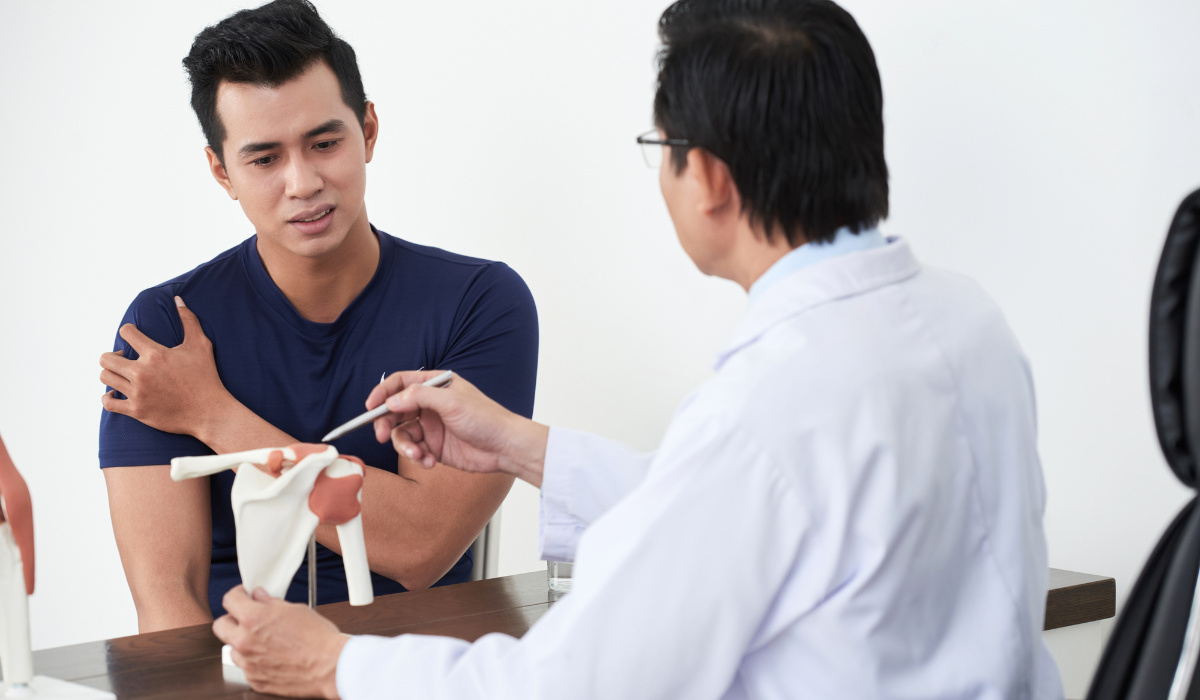
Shoulder arthroscopy is a minimally invasive surgical technique used to diagnose and treat various shoulder joint problems. Through tiny incisions, a small camera (arthroscope) is inserted into the shoulder joint, allowing orthopedic surgeons to view the internal structures on a monitor and perform precise surgical procedures using micro-instruments. This technique has revolutionized shoulder care by offering targeted treatment with minimal disruption to surrounding tissues.
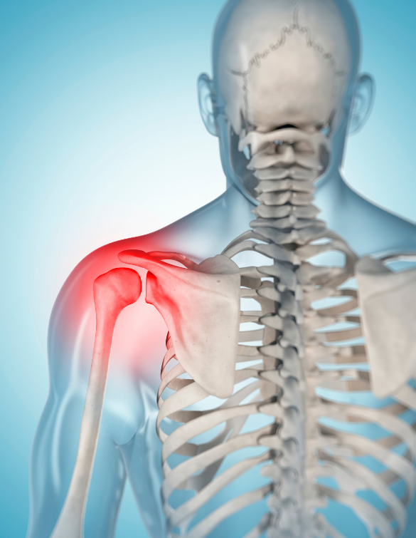
This procedure is commonly used to treat rotator cuff tears, labral tears, shoulder impingement, frozen shoulder, recurrent dislocations, bursitis, and arthritis-related damage. Because it allows for direct visualization of the joint, shoulder arthroscopy provides both accurate diagnosis and treatment in a single procedure. It’s especially suitable for athletes, active individuals, and anyone who requires quicker recovery and minimal downtime.
Benefits of Shoulder Arthroscopy:
Effective, accurate, and less invasive treatment for shoulder conditions.
-
Minimally Invasive Approach
Small incisions reduce muscle trauma, minimize blood loss, and promote faster healing.
-
Faster Recovery Time
Most patients return to light activities within weeks and full function in a few months.
-
Accurate Joint Diagnosis
Direct visualization of the joint enables precise identification and correction of shoulder problems.
Services Offered
We offer comprehensive diagnostic and therapeutic services using advanced shoulder arthroscopy techniques. Our orthopedic specialists begin with a thorough clinical assessment, imaging (X-ray/MRI), and diagnosis to determine if arthroscopy is appropriate for your condition. Once confirmed, the procedure is performed using the latest arthroscopic equipment in a sterile, high-precision environment.
Our arthroscopic procedures address a range of shoulder issues including rotator cuff repair, labrum repair, AC joint arthritis treatment, impingement syndrome relief, subacromial decompression, and removal of loose fragments. Each step is performed using tiny instruments through portals, with real-time visualization to ensure accuracy and joint preservation. The surgery typically lasts 30–90 minutes depending on the condition.
Frequently Asked Questions
To help you better understand your condition and treatment options, we’ve answered some of the most commonly asked questions below.
You may feel mild discomfort after surgery, but pain is well-managed with medication and improves quickly.
Initial recovery is usually 2–6 weeks, but full recovery, including physiotherapy, may take 2–4 months.
Only a few small stitches or skin tapes are used, and they are often removed in a week.
Yes, but only after your surgeon clears you—usually 3–4 months post-op with gradual reintroduction.